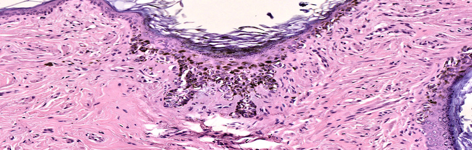
PAGET’S DISEASE. Intraepidermal glandular carcinoma. Dog
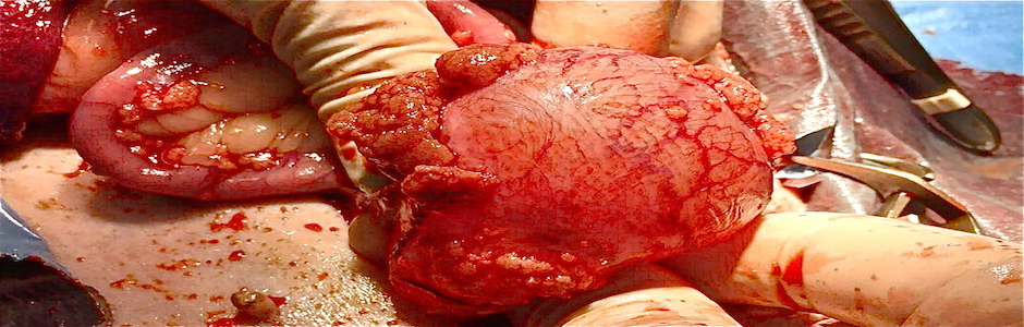
COLLISION TUMOR. Confluence of a carcinoma and osteosarcoma
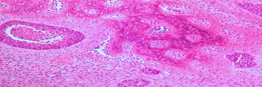
BASAL CELL CARCINOMA. Epithelial neoplasia CK5+ and CK8+
ALOPECIA AREATA. Accumulation of CD4 + and CD8 + lymphocytes in the follicular bulb
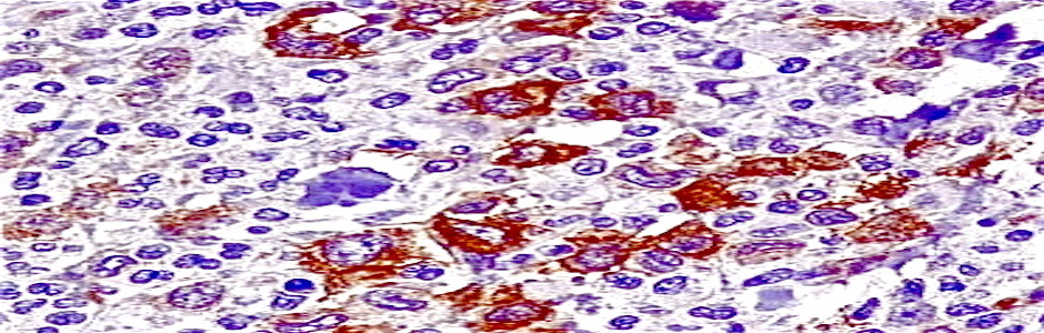
c-kit Type II pattern of expression of c-kit in mast cell tumor. Indicates mutation
MESOTHELIOME. Epithelial neoplasia derived from serosa.
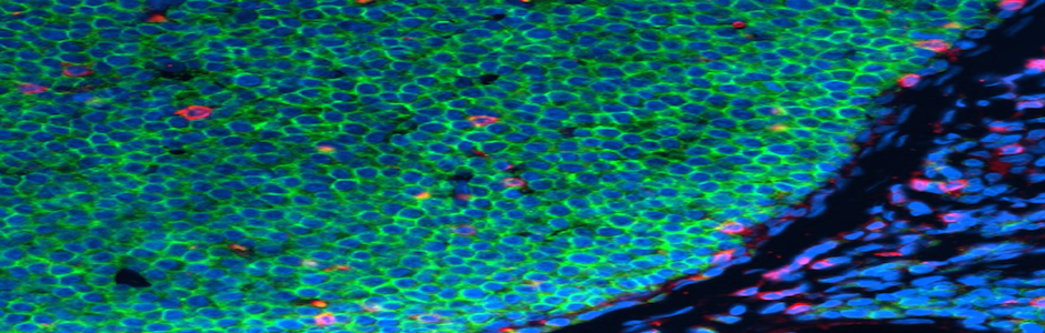
DOUBLE IMMUNOLABELLING B LYMPHOM. Green B lymphocytes (CD79+). Red T lymphocytes (CD3+).
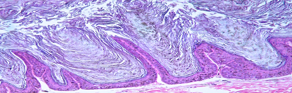
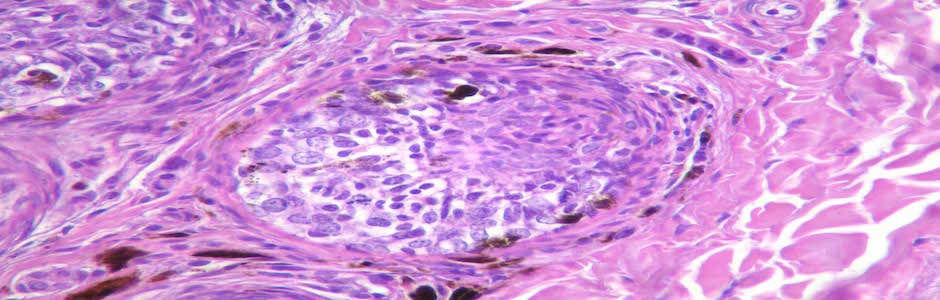
PIGMENTED VIRAL PLAQUE. Papillomavirus infection
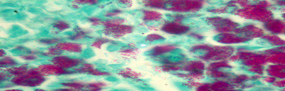
MYCOBACTERIOSIS. Granulomatous lymphadenitis in a Schnauzer associated with Mycobacterium avium subsp. Hominissuis
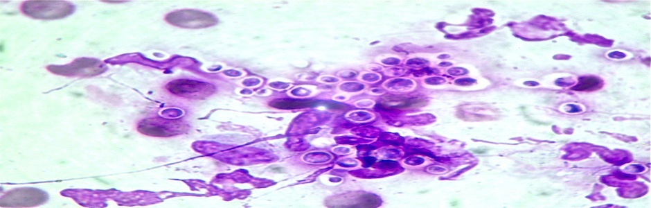
CRYPTOCOCCOSIS. Monomorphic organisms 1-7 µm in diameter with capsular transparent halo
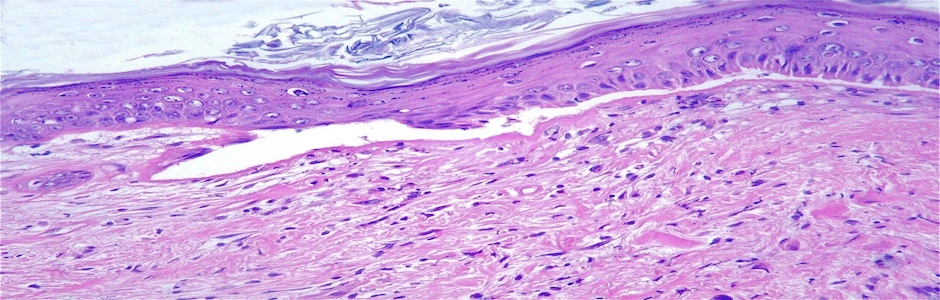
EPIDERMOLYSIS BULLOSA. Separation of the epidermis and dermis
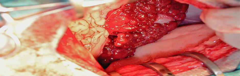
CIRRHOSIS. Fibrosis and nodular hyperplasia
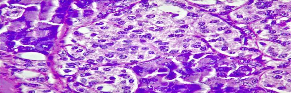
INSULINOMA. Insulin producing beta cells adenoma
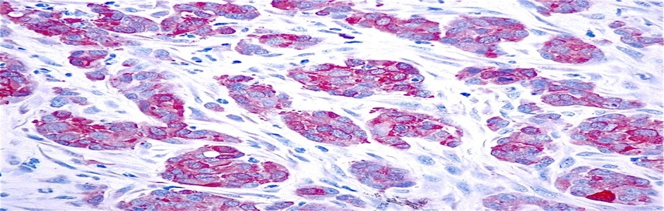
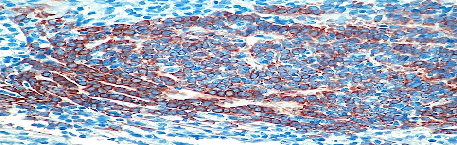
CYTOKERATIN. Cytokeratin positive cells in a carcinoma
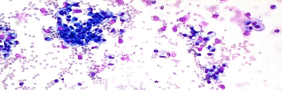
PLEOCYTOSIS. Increased inflammatory cells in the cerebrospinal fluid in encephalitis
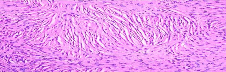
SCHWANOMA. Benign neural tumor
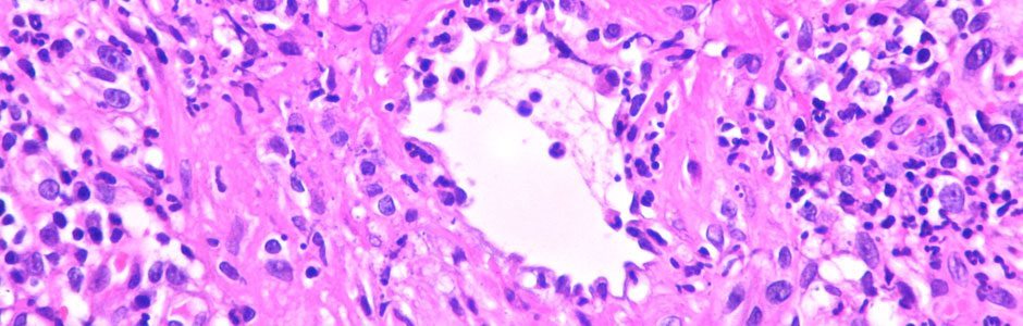
VASCULITIS. Type III hypersensitivity reaction
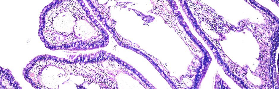
LHYMFAGIECTASIA. Dilated lymph vassels in a protein losing enteropathy